Research Programs
Dr Geddes’s and the IoF’s research is focused at the development of new Fluorescence and Plasmonics concepts and theory, with a view to their downstream biomedical application in health care, safeguard and diagnostics.
Metal-Enhanced Fluorescence (MEF): Metal-Fluorophore Interactions

Figure 1: Metal-Enhanced Fluorescence (MEF) near silver surfaces - significanly increasing detection in fluorescence based systems, top. Bottom: Current mechanistic model for MEF developed by Geddes and co-workers. Click to see larger image.
Although fluorescence is a very highly sensitive technique, where single molecules can routinely be detected, there is still a significant drive for reduced detection limits. The detection limit is usually limited by the quantum yield of the fluorophore, the autofluorescence of the samples and the photostability of the fluorophores. However, there has been a recent explosion in the use of metallic nanostructures to favorably modify the spectral properties of fluorophores and to alleviate some of their photophysical constraints. The use of fluorophore-metal interactions has been termed metal-enhanced fluorescence (MEF)by Dr Geddes and his labs. The uses of MEF have included the increased detectability and photostability of fluorophores, improved protein, DNA / RNA detection, the release of self-quenched fluorescence of over labeled proteins, enhanced wavelength-ratiometric sensing and the application of metallic surfaces to amplified assay detection, to name but just a very few. Dr Geddes’s laboratories have also developed many nanostructured surfaces for metal-enhanced fluorescence such as those comprised of silver islands, silver colloids, silver nano triangles, silver nanorods, and even fractal-like silvered surfaces to name but just a very few. Several modes of silver deposition have also been developed, such as silver deposition by light and electrochemically, on glass, plastics and even using electrodes. Figure 1 visually demonstrates the benefits of Metal-enhanced Fluorescence. As the laser excitation source is moved over an equally coated fluorophore-protein labeled silvered surface, enhanced fluorescence can be observed. The enhanced fluorescence is not due to reflected photons from the mirrored surface as the photographs were taken through an emission filter, but is in fact due to plasmon-coupling and amplification. The ability to increase fluorescence signatures, potentially up to several million-fold, is finding profound applications in the biosciences and in microscopy, where photon flux and detectability is a primary concern.
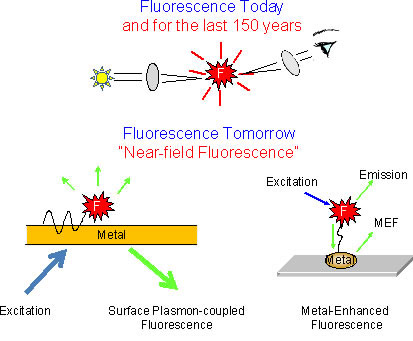
Figure 2: Metal-Enhanced Fluorescence (MEF), A paradigm shift in the way we both use and think about fluorescence spectroscopy. Click to see larger image.
Fluorescence was originally discovered by Sir George Stokes in the 1840s. Since that time we have made notable achievements as a community in understanding all the far-field concepts related to fluorescence. It is for this reason that we have only observed incremental advances in fluorescence and its practices in the last 10 or so years. For example, we can develop new fluorophores with more finely tuned properties, we can develop better instruments and software for fluorescence analysis, but these developments are only incremental, the free space properties and oscillator strength of the fluorophores fundamentally unchanged, with the spatial distribution of the far-field emission remaining for the most part constant. However, Metal-Enhanced Fluorescence (MEF) and indeed plasmonics as a whole, threatens to change the way we both use and think about fluorescence and its applications. In the near-field, fluorophores (dipoles) and metals can couple and favorably modify both near & far-field fluorescence properties. In contrast to typical fluorescence spectroscopy we know very little about near-field fluorescence properties as a community. Subsequently, Dr Geddes’s research is focused at fundamentally understanding these near-field photophysical properties with outcomes focused at translating plasmonic applications into the biosciences. It is Dr Geddes’s current opinion that we are on the verge of a paradigm shift in the uses and utility of Fluorescence spectroscopy, with the next 10 years focusing heavily on new near-field fluorescence concepts, Figure 2.
Progress towards a Unified Plasmon-Fluorophore Theory:
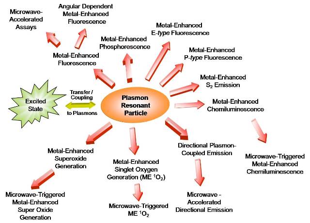
Figure 3: Some Metal-Enhanced Photophysical Concepts developed by the Geddes group (IoF). Click to see larger image.
Over the last 5 or so years, Dr Geddes and the IoF has extensively studied many metal-enhanced photophysical phenomenon. Accordingly, a mechanism for MEF has been postulated. In the current mechanistic interpretation of MEF, non-radiative energy transfer occurs from excited distal fluorophores to the surface plasmon electrons on non-continuous films, in essence an induced mirror dipole. The surface plasmons in-turn, radiate the photophysical characteristics of the coupled quanta. Subsequently, armed with a developed understanding of MEF, research has been intensely focused at both predicting and then experimentally verifying a whole host of metal (plasmon) enhanced photophysical phenomenon, Figure 3, such as Metal-enhanced S2 emission, Metal-enhanced structured fluorescence emission, Metal-enhanced singlet oxygen generation and superoxide generation, and Metal-enhanced multi-photon excitation, to name but just a very few. These collective observations have also led us to postulate the first unified plasmon-fluorophore description, which accounts for many past and present plasmon-dipole observations. At present, this description is still being developed by Geddes and his colleagues, and intense research is likely to continue for several years to come.
Microwave-Accelerated Plasmonics:
Dr Geddes and his research group have recently developed a platform technology for ultra fast and ultra sensitive clinical and bio-agent sensing. The new technology combines the use of Metal-Enhanced Fluorescence (MEF) to amplify fluorescence signatures up to a million-fold, with the use of low power microwaves to kinetically accelerate assays to completion within seconds.. The microwaves are focused at a distance above the nanostructures, by the structures, which is readily modeled using approaches such as Finite-Difference Time Domain, a solution to Maxwell’s equations for propagating EM radiation over complex dielectric substrates / structures. The resultant technology, Microwave-Accelerated Metal-Enhanced Fluorescence (MAMEF), when applied to clinical or bio-agent detection, allows for their fast and sensitive detection. To date, Geddes and his staff have developed ultra fast and sensitive DNA, RNA and protein platforms, such as for Anthrax detection and clinical heart attack markers. This technology may allow first responders to assess on-site, both the nature and concentration of a reagent, without the need for time consuming laboratory processing, such as by real-time PCR. At present there is significant commercial interest in MAMEF. Future research will be focused at applying the technology to high-throughput analysis, such as in HTS plates and a variety of bio warfare or clinical agents simultaneously, i.e. Multiplexing. The current detection limits are about 100 copies of a genome detected within 20 seconds using the MAMEF technology.
Microwave-Accelerated Fluorescence:
While the applications of microwaves have primarily been focused at bulk heating and telecommunications in years past, recent research by Dr Geddes has shown that low power microwaves can have significant benefits in biology also, overcoming some of the classical constraints with protein-protein and DNA-DNA associations. The rapid sampling of binding pockets for species “microwave-accelerated” significantly reduces the extents of non-specific biological absorption. For commonly used practices such as protein or DNA blotting, the Geddes group has shown significant improvements in surface DNA / Protein detectability when employing microwave acceleration. These findings have not only improved the sensitivity of the MAMEF assays (by reduced non-specific absorption), but the notion has lead to the development of the first Microwave-Microscope for the imaging of biological events, published in Optics Express very recently. In addition, Dr Geddes, has undertaken the first Microwave-Accelerated Fluorescence Correlation Spectroscopy (MA-FCS) experiments. This new technology aims to probe microwave accelerated biological events at the single molecule level. Such studies are thought to potentially yield a wealth of biological binding information, not accessible via other approaches. Dr Geddes subsequently aims to develop a MA-FCS instrument in the next several years.
Plasmon Scatter: Theory and Application
Surface plasmons are collective oscillations of free electrons at metallic surfaces. These oscillations can give rise to the intense colors of solutions of plasmon resonance nanoparticles and/or very intense scattering. While the use of plasmonic particle absorption based bioaffinity sensing is now widespread throughout biological research, the use of their scattering properties is relatively ill explored. We refer to the use, utility and control of surface plasmons as Plasmonics.
In this institute of fluorescence new plasmon scattering theory and applications are being developed. These include the use of plasmon scatter for long-range immunosensing and macromolecular conformation studies, as well as the ability to image cell surface receptors for cancer imaging.
Perhaps one of the most profound future applications of plasmon scatter, is likely to be in the measurement of distances in the range 10-300 nm for biological systems. Today, optical distance measurements less than 10 nm are undertaken using FRET between a fluorescent donor and an acceptor. Distances ranging from macroscopic to about 1/2, typically about 300 nm, can be measured using confocal, multiphoton and/or laser scanning methods. While atomic force microscopy and scanning tunneling microscopy can measure these distances, these approaches are not readily compatible with biological species, such as with live cells. However, Dr Geddes is currently investigating the possible use of long-range plasmon coupling between particles to measure such distances, where the coupling occurs to a minimum value at about 2.5 times the diameter of the colloids. Subsequently, long-range FRET based on plasmonics will be possible, based on the changes in the scattering, absorption and polarization properties of suitably sized colloids. Interestingly, the coupling distance (transfer distance in FRET) will be dependent on the wavelength of light and the initial choice of colloid size. This approach is likely to be of significant importance for studying macromolecular dynamics and particularly in immunoassays, which typically have physical dimensions far too large for classical FRET. While the theory for distance measurements using plasmonics has yet to be realized, the lack of a discrete physical dipole may prove simpler than fluorophore dipole-dipole coupling. Theory in this regard is currently underway by Dr Geddes and is likely to find immense importance in the biological and clinical sciences. Several Plasmon scattering papers can be readily observed in Dr Geddes’s publication list. We particularly note a publication in JACS, “Angular-Ratiometric Plasmon-Resonance based Light Scattering for Bioaffinity Sensing” Journal of American Chemical Society, 127(34), 12115-12121, 2005, which describes a new approach to light collection for potential imaging and sensing applications, which is not-prone to system light fluctuations, detector drifts or changes in ambient background light.
Novel-Fluorescence Sensing Concepts:
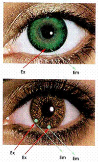
Figure 4: A glucose sensing contact lens for diabetes care and management.
Click to see larger image.
Dr Geddes has developed numerous novel fluorescence sensing methodologies and new fluorescence (and luminescence) probes. Amongst the most highly cited has been the glucose sensing contact lens, Figure 4, a fluorescence-based technology developed by Dr Geddes for diabetes health care monitoring and management. A fluorescent molecule has been developed which readily chelates glucose with high affinity. When embedded in a contact lens, the wearer can readily see changes in the color of the small spot, which tracks the glucose levels in tears and therefore in blood. By comparison with a calibrated color strip the wearer is readily aware of the blood glucose levels and can apply treatment as needed. The technology is completely non-invasive and continuous. Dr Geddes is currently, advancing this program to detect various clinical agents in human tears, such as sodium, potassium, lithium etc, as well as testing the utility of the lens to detect a soldiers / first responders exposure to Bio warfare agents, such as cyanide.
Microwave-Focusing in Biological Systems and with Bioagents:
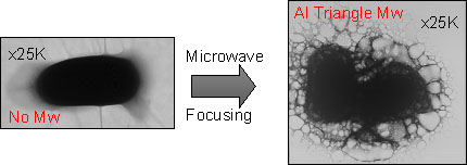
Figure 5: TEM (Transmission Electron Micrograph) images of an Anthrax spore before (left image) and after (right image) being exposed to focused microwaves. The right image shows disruption of the spore, allowing sampling of the genomic DNA using the ultra fast and sensitive MAMEF assay. Click to see larger image.
A research area of growing interest in Dr Geddes’s Institute of Fluorescence is in the use of micro and nanometer size structures for focusing microwave energy. The search for suitable and indeed optimal structures can be modeled using FDTD approaches, and then simply fabricated using thermal vapor deposition techniques. These structures are finding utility in their ability to lyse biological bacterial and cells very rapidly. One recent application in the IoF has been to use intensely focused microwaves to lyse Anthrax spores. Typically, long and relatively expensive digestion protocols, are needed to lyse spores. However, the microwave focusing technology can lyse the spore in under 10 seconds at minimal cost. Given the low power requirements for this approach, it is thought that “field deployable” devices can be developed for the lysing of bacteria in the field, allowing an accurate identification from the released genomic DNA. Figure 5 shows an Anthrax spore, very rapidly lysed by microwave focusing.
International Recognition of the IoF
Under Dr Geddes’s leadership the Institute of Fluorescence receives significant press attention and research coverage. In the last several years, Dr Geddes has given numerous TV interviews, appeared on the Discovery Channel, has appeared in newspapers as well as being reviewed in popular magazines, such as Photonics Spectra, Pharmagenomics, Biophotonics International, to name but just a few. More significantly, Dr Geddes’s research has been highlighted in editorials at the front of several notable journals, including, Nature, JACS, Analytical Chemistry. A list of recent press can be found on the Press section at the IoF Website.
- Metal-Enhanced Fluorescence (MEF)
- Progress towards a Unified Plasmon-Fluorophore Theory
- Microwave-Accelerated Plasmonics
- Microwave-Accelerated Fluorescence
- Plasmon Scatter
- Novel-Fluorescence Sensing Concepts
- Microwave-Focusing in Biological Systems and with Bioagents
- International Recognition of the IoF